


The anatomical structure of the venous system of the lower extremities is characterized by great variability.Knowledge of the individual characteristics of the structure of the venous system plays a large role in evaluating instrumental examination data and choosing the correct treatment method.
The veins of the lower extremities are divided into superficial and deep.The superficial venous system of the lower extremities starts from the venous plexuses of the toes, forming a venous network on the instep and the instep cutaneous arch.From there, it originates from the internal and external marginal veins, entering the large and small saphenous veins, respectively.The great saphenous vein is the longest vein in the body, containing 5 to 10 pairs of valves, with a normal diameter of 3-5 mm.It originates in the lower part of the leg, anterior to the medial epicondyle, and ascends into the subcutaneous tissue of the leg and thigh.In the groin area, the great saphenous vein drains into the femoral vein.Sometimes the great saphenous vein in the thighs and legs can be represented by two or even three trunks.The small saphenous vein begins in the lower part of the leg along its lateral surface.In 25% of cases, it drains into the popliteal vein in the popliteal fossa region.In other cases, the small saphenous vein may rise above the popliteal fossa and drain into the femoral vein, the greater saphenous vein, or into the deep vein of the thigh.
The deep veins of the dorsum of the foot begin with the dorsal plantar veins, draining into the dorsolateral venous arch, from where blood flows into the anterior tibial veins.At the level of the upper third of the leg, the anterior and posterior tibial veins merge to form the popliteal vein, which lies lateral and slightly posterior to the artery of the same name.In the area of the popliteal fossa, the small saphenous vein and the knee joint vein drain into the popliteal vein.The deep femoral vein usually drains into the femoral vein 6-8 cm from the inguinal fold.Above the inguinal ligament, this vessel receives the epigastric vein, a deep vein surrounding the pelvis, and enters the external iliac vein, which merges with the internal iliac vein at the sacroiliac joint.The common pair of iliac veins begins after the confluence of the external iliac and internal iliac veins.The right and left common iliac veins merge to form the inferior vena cava.It is a large vessel without a valve, 19-20 cm long and 0.2-0.4 cm in diameter.The inferior vena cava has apical and visceral branches, through which blood flows from the lower limbs, lower trunk, abdominal organs and small pelvis.
Perforating (communicating) veins connect the deep veins with the superficial veins.Most of them have valves located on the fascia and thanks to which blood moves from the superficial veins to the deep veins.There are direct and indirect perforating veins.Direct lines connect directly with the network of deep and superficial veins, indirect lines connect indirectly, that is, they first flow into the muscle veins, and then into the deep veins.
Most perforating veins arise from branches rather than from the trunk of the great saphenous vein.In 90% of patients, the perforating veins on the inner surface of the lower leg are weakened.In the lower leg, weakness of the perforating Cockett's veins, which connect the posterior branch of the great saphenous vein (Leonardo's vein) with the deep veins, is often observed.In the middle and lower third of the thigh, there are usually 2-4 most permanent perforating veins (Dodd, Gunter), directly connecting the trunk of the great saphenous vein with the femoral vein.With variation of small saphenous varicose veins, poorly communicating veins in the middle, lower part of the leg and in the lateral malleolus area are most often observed.
Clinical course of the disease
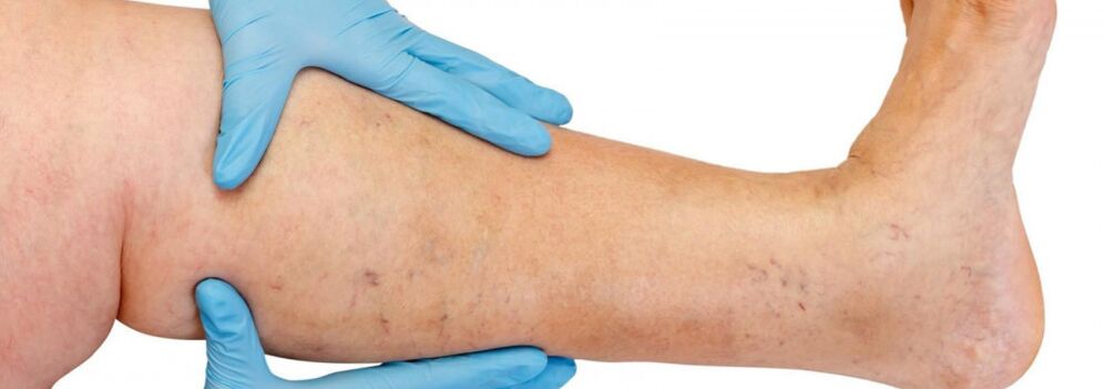
Most often, varicose veins occur in the greater saphenous vein system, less commonly in the small saphenous vein system and begin from branches of the venous trunk in the legs.The natural progression of the disease in the early stages is quite favorable;During the first 10 years or more, apart from cosmetic defects, the patient may not be concerned about anything.Then, if not treated promptly, complaints of heaviness, leg fatigue and swelling after physical activity (walking, standing for a long time) or in the afternoon, especially in the hot season, will begin to appear.Most patients complain of pain in the legs, but when asked for details, it may be revealed that this is precisely a feeling of fullness, heaviness and tightness in the legs.Even with short periods of rest and elevating the position of the limb, the severity of the sensation will decrease.It is these symptoms that characterize venous insufficiency at this stage of the disease.If it comes to pain, other causes need to be excluded (lower limb arterial insufficiency, acute venous thrombosis, joint pain, etc.).Further progression of the disease, in addition to increasing the number and size of varicose veins, also leads to the appearance of trophic disorders, often due to the addition of incompetent perforating veins and the appearance of valvular insufficiency of the deep veins.
In case of lack of perforating veins, trophic disorders are limited to any surface of the leg (lateral, medial, posterior).Nutritional disorders in the early stages are manifested by local hyperpigmentation of the skin, then thickening (hardening) of subcutaneous fatty tissue occurs until cellulite develops.This process ends with the formation of an ulcerative-necrotic defect, which can reach a diameter of 10 cm or more and extend deep into the fascia.The typical location of venous trophic ulcers is the medial malleolus area, but the location of ulcers in the lower legs can vary and vary.In the nutritional disorder stage, the affected area will experience intense itching and burning;Some patients develop bacterial eczema.The pain in the area of the ulcer may not be obvious, although in some cases the pain is very severe.At this stage of the disease, the feeling of heaviness and swelling in the legs becomes constant.
Diagnosis of varicose veins
It is especially difficult to diagnose the preclinical stage of varicose veins, since such a patient may not have varicose veins in the legs.
In such patients, the diagnosis of varicose veins in the legs is mistakenly rejected, despite the presence of symptoms of varicose veins, signs that the patient has relatives with this disease (genetic predisposition), and ultrasound data on early pathological changes in the venous system.
All this can lead to missing the deadline for starting optimal treatment, the formation of irreversible changes in the walls of the veins and the development of very serious and dangerous complications of varicose veins.Only if the disease is detected at an early preclinical stage is it possible to prevent pathological changes in the venous system of the legs through a minimal therapeutic effect on varicose veins.
Avoiding various types of diagnostic errors and making an accurate diagnosis is possible only after a thorough examination of the patient by an experienced specialist, accurate interpretation of all his complaints, detailed analysis of the medical history and maximum possible information about the state of the venous system of the leg obtained using the most modern equipment (specific diagnostic methods).
A duplex scan is sometimes performed to pinpoint the exact site of venous perforation, identifying venous reflux by color coding.In the case of weakened valves, their valves will stop closing completely during the Valsava maneuver or compression test.Valvular insufficiency leads to the occurrence of venous reflux, high, through the incompetent display junction, and low, through the perforating veins in the incompetent leg.Using this method, it is possible to record the backflow of blood through the weakened valve leaflets.That's why diagnosis has many stages or levels.In normal situations, the diagnosis is made after ultrasound diagnosis and examination by a phlebologist.However, in particularly difficult cases, the inspection must be carried out in stages.
- First, a thorough examination and questioning is performed by a phlebologist;
- If necessary, the patient will be sent to additional instrumental research methods (duplex angiography, venography, lymph scintigraphy);
- patients with comorbidities (chondrosis, varicose veins, lymphatic insufficiency) are consulted by leading consultants in these diseases) or additional research methods;
- All patients requiring surgery must first be consulted by a surgeon and an anesthesiologist if necessary.
Treatment
Conservative treatment is indicated mainly for patients with contraindications to surgical treatment: due to the general condition, mild varicose veins cause only cosmetic inconvenience or if surgical intervention is refused.Conservative treatment is aimed at preventing further development of the disease.In these cases, the patient should be advised to bandage the affected surface with an elastic bandage or wear elastic socks, periodically put the leg in a horizontal position and perform special exercises for the foot and leg (flexion and extension at the ankle and knee joints) to activate the muscle-venous pump.Elastic compression accelerates and enhances blood flow in the deep veins of the thighs, reduces the amount of blood in the saphenous vein, prevents the formation of edema, improves microcirculation and helps normalize metabolic processes in tissues.Bandaging should begin in the morning, before getting out of bed.The bandage is applied with light tension from the toes to the thighs, with mandatory adhesion of the heel and ankle joints.Each subsequent bandage loop must overlap half of the previous one.It is recommended to use certified medical hosiery with an individual choice of compression level (from 1 to 4).Patients should wear comfortable shoes with hard soles and low heels, avoid standing for long periods of time, do heavy manual labor, and work in hot and humid places.If due to the nature of work the patient has to sit for a long time, he should place his feet in a high position by placing a special stand of the required height under the feet.It is recommended to walk a little every 1-1.5 hours or tiptoe 10-15 times.The resulting contraction of the calf muscles improves blood circulation and increases venous flow.While sleeping, your legs should be placed in an elevated position.
The patient should limit water and salt intake, normalize body weight and periodically take diuretics and drugs that improve venous tone.According to indications, the drug is prescribed to improve microcirculation in tissues.For treatment, nonsteroidal anti-inflammatory drugs should be used.
Physical therapy plays an important role in preventing varicose veins.For uncomplicated forms, water procedures are useful, especially swimming, foot baths in warm water (not higher than 35°) with a 5-10% solution of table salt.
Compressive sclerotherapy
Indications for injection therapy (sclerotherapy) for varicose veins are still debated.This method involves introducing a sclerosing agent into the varicose vein, causing it to further compress, damage and harden.Modern drugs used for these purposes are quite safe, i.e. do not cause necrosis of the skin or subcutaneous tissue when administered extravascularly.Some experts use sclerotherapy for most forms of varicose veins, while others completely reject it.Most likely, the truth lies somewhere in the middle, and it makes sense for young women in the early stages of the disease to use injection treatments.The only thing is that they must be warned about the possibility of recurrence (higher than with surgical intervention), having to continuously wear a fixed compression bandage for a long time (up to 3-6 weeks) and the possibility of needing several treatment sessions to completely harden the veins.
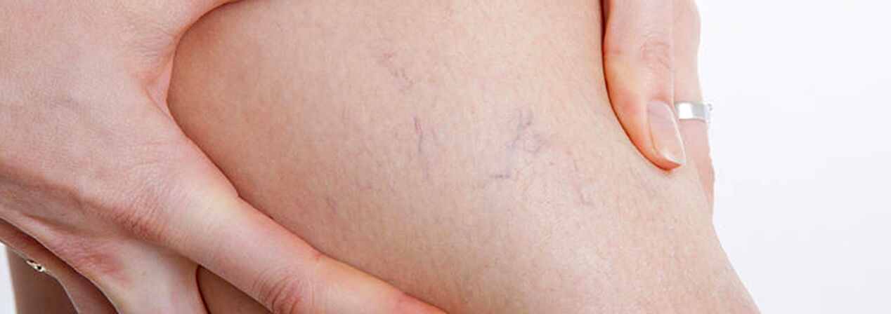
The group of patients with varicose veins should include patients with telangiectasia (“spider veins”) and small saphenous reticular veins, because the causes of development of these diseases are similar.In this case, along with sclerotherapy, you can alsoPercutaneous laser coagulation, but only after excluding damage to deep and penetrating veins.
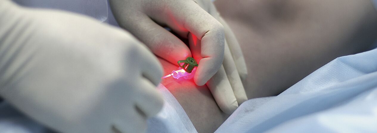
Percutaneous laser coagulation (PLC)
This method is based on the principle of selective photocoagulation (photothermolysis), which is based on the different absorption of laser energy by different substances in the body.The special feature of the method is the non-contact nature of this technology.The concentrator head focuses energy on the blood vessels in the skin.Hemoglobin in the circuit selectively absorbs laser beams of certain wavelengths.Under the influence of laser, destruction of the endothelium occurs in the vessel lumen, leading to adhesion of the vessel wall.
The effectiveness of PLK directly depends on the penetration depth of laser radiation: the deeper the circuit, the longer the wavelength, therefore PLK has quite limited indications.For vessels over 1.0-1.5 mm in diameter, microsclerotherapy is most effective.Considering the widespread distribution and branching of spider veins on the legs and the variable diameter of the blood vessels, a combined treatment method is now actively used: at the first stage, sclerotherapy of veins with a diameter of more than 0.5 mm is performed, then a laser is used to remove the remaining “stars” of smaller diameter.
The procedure is practically painless and safe (no skin cooling drugs and anesthesia are used), because the device's light belongs to the visible part of the spectrum, and the light wavelength is designed so that the water in the tissues does not boil and the patient does not get burned.For patients with high pain sensitivity, it is recommended to use a cream with a local anesthetic effect beforehand.Redness and swelling subside within 1-2 days.After the course, for about two weeks, some patients may experience darkening or lightening of the treated skin area, which then disappears.In people with fair skin, changes are hardly noticeable, but in patients with dark or strongly tanned skin, the risk of such temporary pigmentation is quite high.
The number of procedures depends on the complexity of the case - the blood vessels are at different depths, the lesions can be small or occupy a fairly large surface of the skin, but usually no more than four sessions of laser therapy are needed (5-10 minutes each).Maximum results are achieved in such a short time thanks to the unique “square” shape of the device's light pulses;it increases its effectiveness compared to other devices, while also reducing the likelihood of post-procedure side effects.
Surgical treatment
Surgery is the only radical treatment method for patients with varicose veins of the lower limbs.The purpose of this operation is to eliminate the pathogenic mechanism (venous-venous reflux).This is achieved by removing the main trunks of the greater and lesser saphenous veins and ligation of poorly communicating veins.
Surgical treatment of varicose veins has a history of hundreds of years.In the past, and many surgeons still do, large incisions were used along the varicose veins and general or spinal anesthesia was used.The mark after such a “minor phlebotomy” remains a lifelong reminder of the surgery.The first vein surgeries (according to Schade, according to Madelung) were so traumatic that the harm from them was greater than the harm from varicose veins.
In 1908, American surgeon Babcock devised a method of pulling veins under the skin using a hard metal probe shaped like an olive.In its more improved form, this surgical method of removing varicose veins is still used in many public hospitals.Varicose veins are removed with separate incisions, as suggested by surgeon Narat.Therefore, the classic phlebotomy is called the Babcock-Narat method.Phlebectomy according to Babcock-Narat has disadvantages - large scars after surgery and impaired skin sensitivity.The ability to work is reduced for 2-4 weeks, making it difficult for patients to agree to surgical treatment of varicose veins.
Phlebologists have developed a unique technology to treat varicose veins in one day.Complicated cases are surgically usedcombined technology.The main large varicose veins are removed using the reverse peeling method, requiring minimal intervention through small incisions (from 2 to 7 mm) in the skin, leaving virtually no scars.The use of a minimally invasive technique involves minimal tissue trauma.The result of this operation is the elimination of varicose veins with excellent cosmetic results.Combined surgical treatment is performed under general or intravenous anesthesia, with a maximum hospital stay of up to 1 day.

Surgical treatment includes:
- Transverse - crosses where the trunk of the great saphenous vein flows into the deep venous system;
- Stripping is the removal of a segment of varicose veins.Only varicose veins are removed, not all veins (as in the classic version).
Actuallysmall vein removalhas replaced the Narat technique for removing dilated branches of main veins.Previously, skin incisions from 1-2 to 5-6 cm were made along the line of the varicose veins, through which the veins were separated and removed.The desire to improve the cosmetic results of the intervention and be able to remove veins not through traditional incisions but through small incisions (injections), forced doctors to develop tools that allowed them to do almost the same thing through a minimal skin defect.This is how phlebotomy “hooks” of various sizes and configurations as well as special spoons appeared.And instead of ordinary scalpels, scalpels with very narrow blades or needles with a fairly large diameter began to be used to penetrate the skin (for example, a needle used to take venous blood for analysis has a diameter of 18G).Ideally, puncture marks with such a needle will be practically invisible after a while.
Some forms of varicose veins are treated on an outpatient basis under local anesthesia.Minimal trauma during small vein ablation as well as low risk of intervention allows this surgery to be performed in a day hospital.After minimal observation in the post-operative clinic, the patient can go home on his own.In the postoperative period, an active lifestyle is maintained, active walking is encouraged.Temporary incapacity for work usually lasts no more than 7 days, after which work can begin.
When is microangiectomy used?
- When the diameter of the dilated body of the greater or lesser saphenous vein is greater than 10 mm;
- After thrombophlebitis of the main subcutaneous trunks;
- After recanalization of the trunks after other treatments (EVLT, sclerotherapy);
- Eliminate very large individual varicose veins.
It can be a stand-alone operation or a component of a combination treatment of varicose veins, combined with laser vein treatment and sclerotherapy.The tactics of use are determined individually, always taking into account the results of parallel ultrasound scanning of the patient's venous system.Microphlebectomy is used to remove veins in various locations that have been altered for a variety of reasons, including on the face.Professor Varadi from Frankfurt developed his convenient instruments and formulated the basic principles of modern microangiectomy.Varadi phlebotomy provides excellent cosmetic results without pain or hospitalization.This work is very hard, almost like jewelry making.
After vein surgery
The postoperative period after “classical” phlebectomy is usually quite painful.Sometimes a large hematoma is a concern and swelling occurs.Wound healing depends on the surgeon's surgical technique;Sometimes there is lymphatic leakage and permanent scar formation;Often after a major vein removal surgery, the heel area still loses sensitivity.
On the contrary, after surgery to remove small veins, the wound does not need stitches because it is just a puncture, does not cause pain, and does not actually damage the skin nerves.However, such phlebectomy results are only achieved by very experienced phlebotomy surgeons.